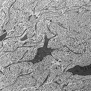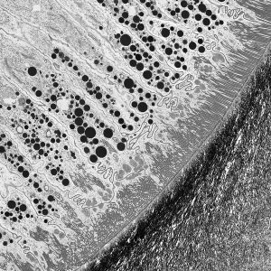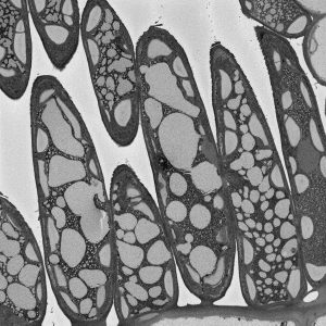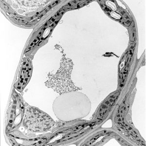

Kathryn Green on a TEM at the University of Queensland. Kathryn took the image of a fig tree leaf.
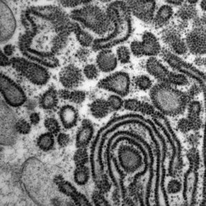
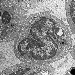
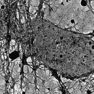
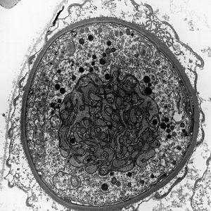
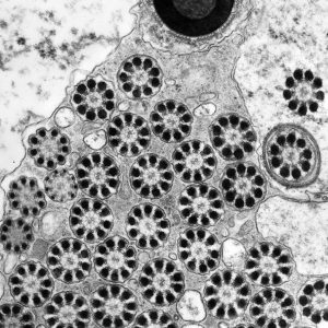
Microscopes are tools that have evolved from the earliest days of small hand-held glass lenses into large and sophisticated instruments that use light, electron and ion beams to reveal some of the deepest secrets of living things and the detailed structures of the materials that make up our world. Inside the microscope, light or electron beams either scan over the surface of a sample or shine through the sample to produce images that hold the stories to be read. The micrographs in this exhibition were created on transmission electron microscopes (TEMs) where an electron beam passes through a very thin slice of the sample being examined. This gives a flat-looking image, rather like an aerial view of the sample. Read more about the microscopy below.
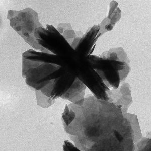
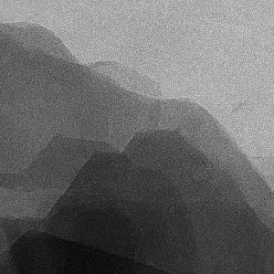
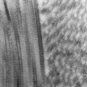
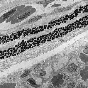
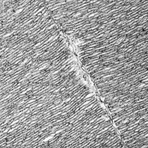
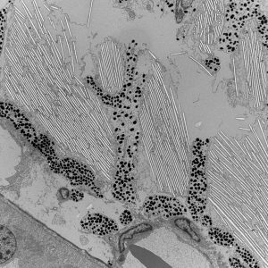
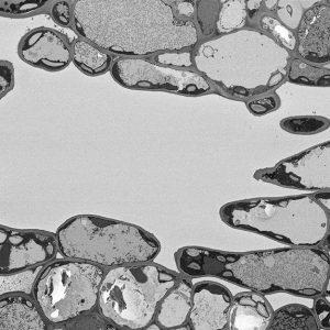
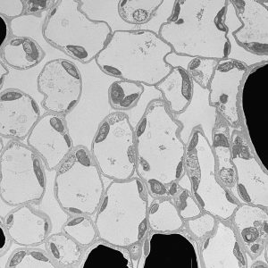
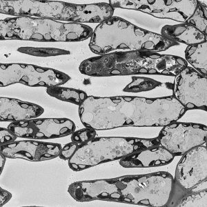
The micrographs in this exhibition were created on transmission electron microscopes (TEMs) where an electron beam passes through a very thin slice of the sample being examined. This gives a flat-looking image, rather like an aerial view of the sample. Where there are denser parts, the electron beam is blocked and a dark area is created on the image. Where there is less material, the beam goes through easily and shows up as a lighter area on the image. These images can only be captured in black and white as no visible light is involved in the imaging process.
Microscopes allow us to look deep into all manner of things such as ancient rocks, cells from living things and high-tech new materials. They reveal information that can lead to new treatments for disease, new ways to protect and repair our environment, and better materials for us to build new and more environmentally sensitive and efficient technologies. Microscopy also supports industry. For example, it helps to keep rescue helicopters flying safely by providing information for forensic deduction. Microscopy Australia works with CHC Helicopter to review oil samples and quickly identify if there are any metal filings in a sample and if so, to identify the type of metal. CHC can then identify which component part the metal may have come from and what they need to do to fix it. This is just one example of how microscopy is at work behind the scenes of our lives.
Microscopy Australia is Australia’s national research infrastructure for microscopy. Based in universities around the country, it enables all Australian researchers to access the instruments and expertise they need. All the micrographs in this exhibition were captured on Microscopy Australia instruments in labs around the country.