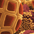
Our advanced microscopes can capture movement and 3-dimensional data on video. This helps scientists learn from the natural world and design new technologies.
Learn more about the chiton teeth in this 3D data.
Visualised using X-ray microtomography by Dr Jeremy Shaw, University of Western Australia and A/Prof. Allan Jones, University of Sydney.
Size: the green area is about 1 millimetre wide.
Learn more about the metal alloy in this 3D data.
Visualised using atom probe tomography by Dr Gang Sha and Dr Baptiste Gault, University of Sydney.Size: sample is 150 nanometres high.
Read about this soil flythrough.
Visualised using X-ray microtomography by Tim Burykin and Prof. John Crawford at the University of Sydney. The whole sample is 1 cubic millimetre in size.
Read about muscle structures in this 3D data.
Visualised using electron tomography by Garry Morgan, Charles Ferguson and Prof. Rob Parton, University of Queensland. Size: each caveola is approximately 50 nanometres in diameter.
Read about the leaf structures in this 3D data.
Visualised using X-ray microtomography by Tim Burykin, Prof. John Crawford, A/Prof. Margaret Barbour and Elinor Goodman, University of Sydney. Size: the scanned areas are both 220 micrometres wide.
Read about breast cancer cell movement captured in this video.
Visualised using differential interference microscopy by Dr Sandra Fok and Dr Lilian Soon, University of Sydney. Size: the field of view is 70 micrometres wide.
Read about this spider web attachment complex.
Visualised using 3D electron microscopy by Dr Jonas Wolff, Macquarie University and Dr Minh Huynh, University of Sydney. Size: the attachment complex is approximately 300 micrometres in diameter.
Read about the 7000,000 year old ant in this 3D X-ray data.
Visualised using X-ray microtomography by A/Prof. Allan Jones, University of Sydney. Size: the ant is 3.8 mm long

